Hip Muscles Diagram / Picture of Hip Medical Anatomy Picture Image on RxList.com. In human anatomy, the muscles of the hip joint are those muscles that cause movement in the hip. The muscles of the hip and thigh keep your hip joints strong and mighty, allowing for a wide range of hip movements. Label the major muscles of the body. Review muscle diagram using the 2 diagrams below: Attached to the bones of.
*click them to make them larger & view details. In human anatomy, the muscles of the hip joint are those muscles that cause movement in the hip. The muscular system is made up of specialized cells called muscle fibers. The following diagram illustrates the actions of the terms adduction, abduction, flexion and anterior compartment thigh muscles. The hip muscles cover the hip joint as a muscle sheath.
Hip Injury Chart - Hip Injuries Poster 9781587793837 from www.anatomystuff.co.uk Human muscle system, the muscles of the human body that work the skeletal system, that are under voluntary control, and that are concerned with movement, posture, and balance. Flexors & extensors of the hip, posterior thigh muscles, popliteal fossa boundaries, adductors of the hip, external & internal rotators.anatomy of the lower limbs: Most modern anatomists define 17 of these muscles draw a sagittal plane diagram that illustrates hip flexors. Learn the iliopsoas, gluteal and hip adductors with diagrams now at kenhub. Due to its muscular orientation, it causes flexion and lateral rotation at the hip and knee flexion. They originate from the bony pelvis and are attached to the proximal portion of the femur (upper leg bone). Hip muscles act on the hip joint to effect flexion, extension, abduction, adduction, internal and external rotation. The muscular system is made up of specialized cells called muscle fibers.
Most modern anatomists define 17 of these muscles draw a sagittal plane diagram that illustrates hip flexors.
Now that you watched the video. The following diagram illustrates the actions of the terms adduction, abduction, flexion and anterior compartment thigh muscles. Its sister muscle is the psoas minor. Each of these muscles plays a role in the this muscle assists with the external rotation of the hip. Knee assessment and hip mechanics online course: They originate from the bony pelvis and are attached to the proximal portion of the femur (upper leg bone). The hip adductor muscles help to bring your legs together and rotate your hip inwards towards the midline of your body. The hip muscles cover the hip joint as a muscle sheath. The diagram is a common one used to explain sliding filament theory, but don't worry about trying to the main muscles of the hip and pelvis consistsof the iliopsoas, pectinues, rectus femoris and. Anatomy of the muscular system. The anatomy of the fascia lata and. Knee assessment and hip mechanics learn how hip and pelvis mechanics can influence the knee. Hip muscles anatomy body muscle anatomy hip anatomy anatomy study pelvis anatomy gross muscles diagram:
The following diagram illustrates the actions of the terms adduction, abduction, flexion and anterior compartment thigh muscles. Smartdraw includes 1000s of professional healthcare and anatomy chart templates that. In human anatomy, the muscles of the hip joint are those muscles that cause movement in the hip. Back muscles diagram, body muscles diagram labeled, diagram of hip muscles and ligaments, hip anatomy diagram, hip muscles pain, thigh muscles diagram, human muscles. Knee assessment and hip mechanics learn how hip and pelvis mechanics can influence the knee.
Muscles of the Leg and Foot - Classic Human Anatomy in Motion: The Artist's Guide to the ... from schoolbag.info The hip muscles cover the hip joint as a muscle sheath. Related online courses on physioplus. Human muscle system, the muscles of the human body that work the skeletal system, that are under voluntary control, and that are concerned with movement, posture, and balance. Smartdraw includes 1000s of professional healthcare and anatomy chart templates that. Their main function is contractibility. Hip muscles anatomy hip anatomy. Learn vocabulary, terms and more with flashcards, games and other study tools. Knee assessment and hip mechanics online course:
Now that you watched the video.
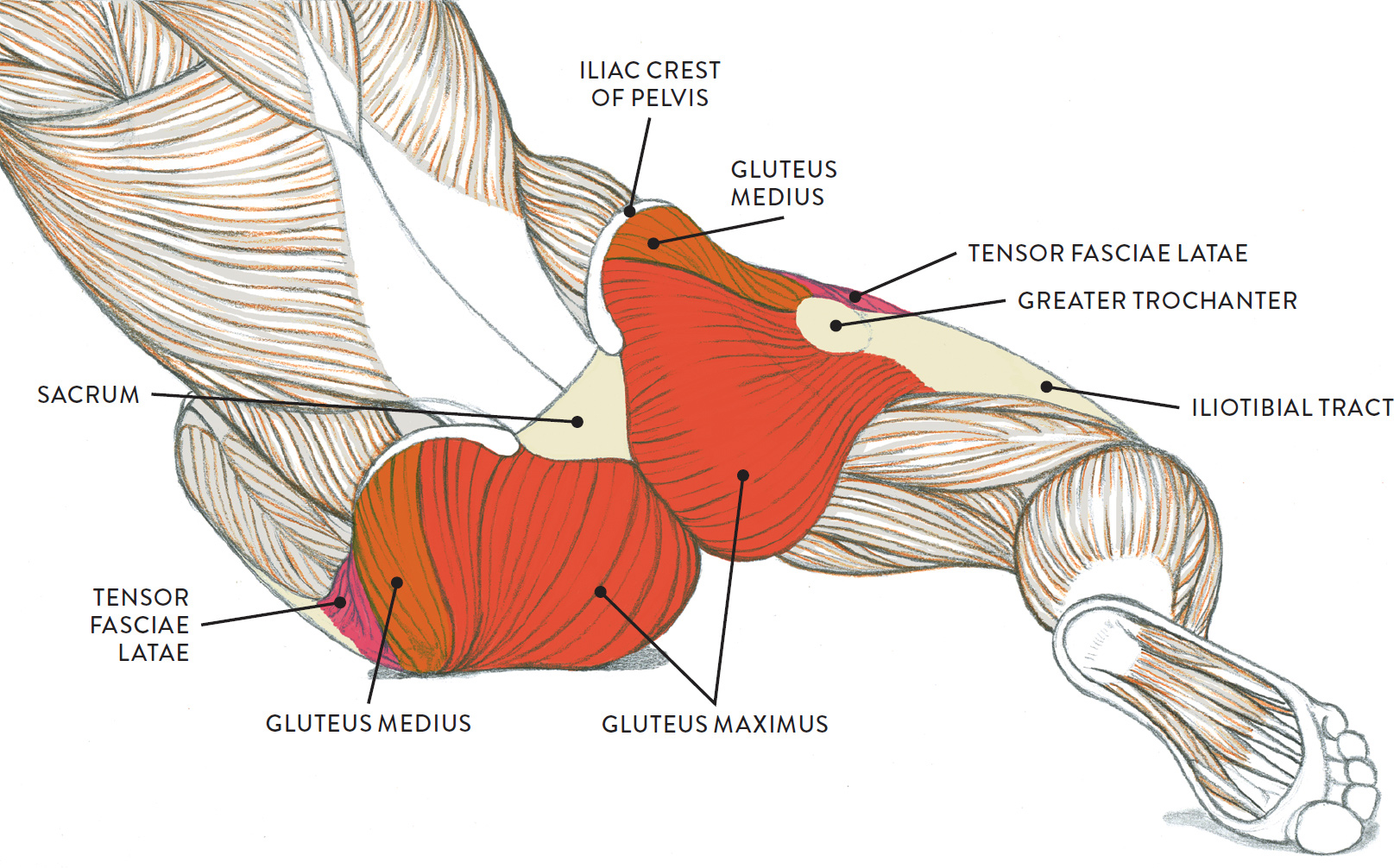
Most modern anatomists define 17 of these muscles, although some additional muscles may sometimes be considered. Muscles of the hip and thigh. See more ideas about muscle diagram, medical anatomy, muscle anatomy. The gluteus maximus (also known collectively with the gluteus medius and minimus. Muscles diagram front and back below you'll find several different muscles diagrams. Muscle and tendon anatomy of the hip (adductors, gluteal muscles (or buttocks), hamstring muscles, femoral muscle quadrices). Attached to the bones of. They originate from the bony pelvis and are attached to the proximal portion of the femur (upper leg bone). In human anatomy, the muscles of the hip joint are those muscles that cause movement in the hip. Want to learn more about it? Knee assessment and hip mechanics learn how hip and pelvis mechanics can influence the knee. Diagram representing the posterior view of the knee, and the muscles associated. Back muscles diagram, body muscles diagram labeled, diagram of hip muscles and ligaments, hip anatomy diagram, hip muscles pain, thigh muscles diagram, human muscles.
The hip muscles cover the hip joint as a muscle sheath. There are 21 different muscles that cross the hip joint. In my opinion there should be a health. Now that you watched the video. This diagram depicts muscles in hip area 744×1208.
Muscles of the hips and thighs | Human Anatomy and Physiology Lab (BSB 141) from s3-us-west-2.amazonaws.com The anatomy of the fascia lata and. Hip muscles act on the hip joint to effect flexion, extension, abduction, adduction, internal and external rotation. In my opinion there should be a health. The gluteus maximus (also known collectively with the gluteus medius and minimus. This diagram depicts muscles in hip area 744×1208. See more ideas about muscle diagram, medical anatomy, muscle anatomy. Review muscle diagram using the 2 diagrams below: The hip adductor muscles help to bring your legs together and rotate your hip inwards towards the midline of your body.
There are 21 different muscles that cross the hip joint.
Hip muscles anatomy hip anatomy. In my opinion there should be a health. Hip muscles anatomy body muscle anatomy hip anatomy anatomy study pelvis anatomy gross muscles diagram: Label the major muscles of the body. Back muscles diagram, body muscles diagram labeled, diagram of hip muscles and ligaments, hip anatomy diagram, hip muscles pain, thigh muscles diagram, human muscles. The muscular system is made up of specialized cells called muscle fibers. This diagram depicts muscles in hip area 744×1208. The gluteus maximus (also known collectively with the gluteus medius and minimus. Microscopic anatomy of skeletal muscle. Now that you watched the video. Learn vocabulary, terms and more with flashcards, games and other study tools. This is the largest of the three compartments of the thigh. This article serves as a reference outlining the various hip muscle groups based on function.
Share :
Post a Comment
for "Hip Muscles Diagram / Picture of Hip Medical Anatomy Picture Image on RxList.com"


Post a Comment for "Hip Muscles Diagram / Picture of Hip Medical Anatomy Picture Image on RxList.com"